

Fibronectin is a high - molecular - weight glycoprotein that plays a crucial role in various biological processes, such as cell adhesion, migration, growth, and differentiation. As a Fibronectin supplier, I've been really interested in what factors can regulate its expression. Understanding these factors can not only help us better understand the biological functions of Fibronectin but also guide us in optimizing its production and application.
1. Growth Factors and Cytokines
Growth factors and cytokines are like the messengers in our body, sending signals to cells to regulate different biological activities. They have a significant impact on Fibronectin expression.
Transforming Growth Factor - beta (TGF - β) is a well - known regulator. TGF - β can stimulate Fibronectin synthesis in many cell types, including fibroblasts, endothelial cells, and epithelial cells. When TGF - β binds to its receptors on the cell surface, it activates a series of intracellular signaling pathways, such as the Smad pathway. This activation leads to the up - regulation of Fibronectin gene transcription. For example, in wound healing, TGF - β is released at the injury site. It promotes the production of Fibronectin by fibroblasts, which helps in the formation of the extracellular matrix (ECM) and the migration of cells to the wound area.
Platelet - Derived Growth Factor (PDGF) also affects Fibronectin expression. PDGF is released by platelets during the early stages of wound repair. It can increase the synthesis of Fibronectin in fibroblasts. The binding of PDGF to its receptors activates tyrosine kinase activity, which in turn activates downstream signaling pathways. These pathways can enhance the expression of Fibronectin, contributing to cell proliferation and migration during tissue repair.
Interleukin - 1 (IL - 1) is a cytokine that can have both positive and negative effects on Fibronectin expression, depending on the cell type and the context. In some cases, IL - 1 can up - regulate Fibronectin production in fibroblasts. However, in other cell types, it may have an inhibitory effect. This complex regulation shows that the interaction between cytokines and Fibronectin expression is highly context - dependent.
2. Hormones
Hormones are another group of important regulators of Fibronectin expression.
Estrogen has been shown to influence Fibronectin synthesis. In female reproductive tissues, such as the uterus and breast, estrogen can increase the expression of Fibronectin. Estrogen binds to its receptors in the cell nucleus, and the hormone - receptor complex then binds to specific DNA sequences, called estrogen - response elements (EREs), in the Fibronectin gene promoter region. This binding can enhance the transcription of the Fibronectin gene. For instance, during the menstrual cycle, the changing levels of estrogen can lead to corresponding changes in Fibronectin expression in the uterine endometrium, which is important for embryo implantation.
Glucocorticoids, on the other hand, can have an inhibitory effect on Fibronectin expression. Glucocorticoids are stress hormones that are released in response to various stressors. They bind to glucocorticoid receptors in the cell, and the complex can interact with transcription factors involved in Fibronectin gene regulation. This interaction often leads to a decrease in Fibronectin synthesis. In some inflammatory diseases, the use of glucocorticoid drugs can reduce the excessive production of Fibronectin, which may be beneficial in preventing fibrosis.
3. Mechanical Stress
Cells in our body are constantly exposed to mechanical forces, and these forces can regulate Fibronectin expression.

In the cardiovascular system, endothelial cells are subjected to shear stress due to blood flow. Shear stress can increase the expression of Fibronectin in endothelial cells. The cells sense the mechanical force through mechanosensors on their surface, such as integrins. When integrins are activated by shear stress, they transmit signals into the cell, which can lead to the up - regulation of Fibronectin gene expression. This increased Fibronectin production helps in maintaining the integrity of the endothelial layer and the proper function of blood vessels.
In the musculoskeletal system, mechanical loading on bone and muscle cells can also affect Fibronectin expression. For example, during exercise, the mechanical stress on muscle fibers can stimulate the production of Fibronectin by muscle satellite cells. Fibronectin then plays a role in muscle repair and regeneration by promoting cell adhesion and migration.
4. Nutrients and Small Molecules
Certain nutrients and small molecules can influence Fibronectin expression.
Vitamin C is essential for the synthesis of Fibronectin. It acts as a co - factor for the enzymes involved in the hydroxylation of proline and lysine residues in Fibronectin. Hydroxylation is important for the proper folding and stability of Fibronectin. Without sufficient vitamin C, the synthesis of functional Fibronectin is impaired. For example, in scurvy, a disease caused by vitamin C deficiency, the lack of proper Fibronectin synthesis can lead to problems in wound healing and connective tissue integrity.
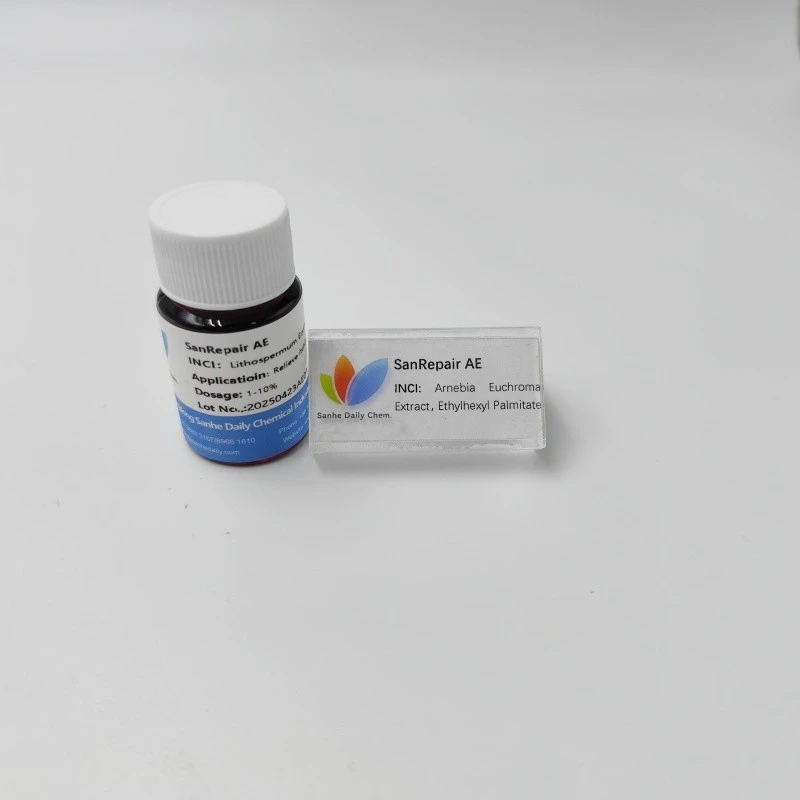
Some small molecules found in natural extracts can also regulate Fibronectin expression. Ceramide NP is a lipid molecule that has been shown to have an impact on Fibronectin production in skin cells. It can enhance the expression of Fibronectin, which is beneficial for maintaining the structure and function of the skin's extracellular matrix. Zinc Oxide is another compound that can influence Fibronectin synthesis. Zinc is an essential trace element, and zinc oxide can promote Fibronectin production in fibroblasts, which is important for wound healing and tissue repair. Lithospermum Erythrorhizon Extract has also been reported to have regulatory effects on Fibronectin expression. It may contain bioactive compounds that can modulate the signaling pathways involved in Fibronectin synthesis.
5. Cell - Cell and Cell - Matrix Interactions
Cell - cell and cell - matrix interactions are crucial for regulating Fibronectin expression.
Integrins are cell - surface receptors that mediate the interaction between cells and the extracellular matrix. When integrins bind to Fibronectin in the ECM, they can activate intracellular signaling pathways. These pathways can either up - regulate or down - regulate Fibronectin expression, depending on the specific integrin subtype and the context. For example, the binding of α5β1 integrin to Fibronectin can lead to the activation of focal adhesion kinase (FAK) and subsequent signaling events that can enhance Fibronectin production.
Cell - cell contact also plays a role. In epithelial cells, for instance, tight junctions and adherens junctions between cells can influence Fibronectin expression. When cells are in close contact, they can exchange signals through these junctions, which can regulate the synthesis and secretion of Fibronectin.
Conclusion
As a Fibronectin supplier, understanding the factors that regulate Fibronectin expression is of great importance. By manipulating these factors, we can potentially optimize the production of high - quality Fibronectin. Whether it's through the use of growth factors, hormones, or small molecules, we can explore different strategies to meet the diverse needs of our customers.
If you're interested in purchasing Fibronectin for your research, pharmaceutical, or cosmetic applications, feel free to contact us for more information and to start a procurement discussion. We're always ready to provide you with the best products and services.
References
- Heldin, C. H., & Miyazono, K. (1993). TGF - β signal transduction. Cell, 75(5), 593 - 596.
- Ross, R. (1986). The pathogenesis of atherosclerosis - an update. New England Journal of Medicine, 314(8), 488 - 500.
- Ingber, D. E. (2003). Mechanobiology and diseases. Journal of Internal Medicine, 254(6), 564 - 577.
- Prockop, D. J., & Kivirikko, K. I. (1995). Collagens: molecular biology, diseases, and potentials for therapy. Annual Review of Biochemistry, 64(1), 403 - 434.